Process development/ Process control (PAT, cGMP), Production platform for proteins and virus-like particles

Project description:

For a translation of research results from the DRUID consortium into the clinic or industry, robust production processes, which represent more than just scaling up, are essential. These processes must follow the guidelines of good manufacturing practice (cGMP) and process analytical technology (PAT). The following work is planned: i) process intensification and expansion of the BEVS production platform as well as implementation of a continuous process control, ii) integration of online PAT technology, e.g., impedance spectroscopy for an automated determination of the optimal point in time for harvesting and automated harvesting, iii) expression (BEVS) and purification of S. mansoni kinases as well as study of the kinases and putative inhibitors, and iv) study of novel transfection reagents for transient protein production on a bioreactor scale.

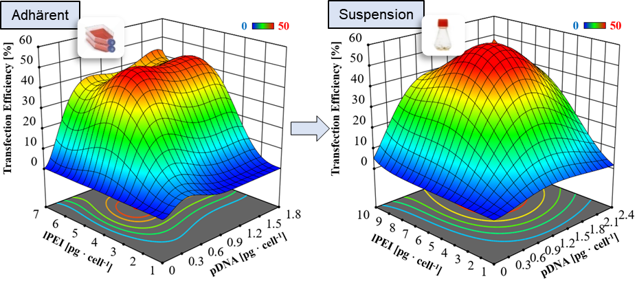
Optimization of the transient transfection process using statistical design of experiments. ©IBPT

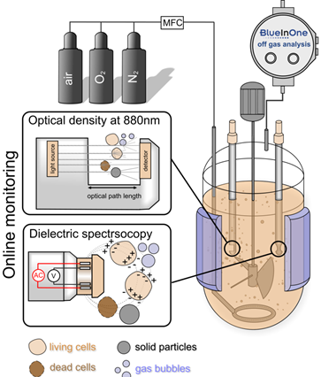
Production concept with integrated PAT technology. ©IBPT


Scientific goal:

The work on production process development and process control will be further adapted to the questions of the center, expanded, and offered to the entire consortium for use. It is planned to further intensify and automate the established BEVS (Baculovirus Expression System) platform. In addition, the second production platform – the transient production with HEK-293T cells – is also being further developed.


DRUID Collaboration partners:

B4 Schlitzer Lab, B5 Grevelding lab, B7 P Falcone Lab, C6 NWG Häberlein, E1 Platform Grevelding/ Häberlein


References E5: Biotechnol, DOI: 10.1016/j.ejbt.2021.08.002, 3. Lothert et al. (2020) Methods Mol Biol 2183:217-248, 4. Dekevic et al. (2022) J Biotechn 346 23-34, 5. Schwarz et al. (2021) Elec J Biotechnol, DOI: 10.1016/j.ejbt.2022.01.003; 6. Barekzai et al. (2020) New Adv Ferm Processes, DOI: 10.5772/intechopen.90029, 7. Eckhardt et al. (2021) Sep Sci Technol, 57 (6) 886-897

High resolution mass spectrometry imaging

Project description:

High resolution mass spectrometry imaging provides numerous opportunities to aid in the characterization of pathogens, host-pathogen interactions and the analysis of drug molecules. The sample is analyzed with a laser in a rasterized fashion, the step size is only a few micrometers. The sample material is ablated and ionized, allowing for the parallel detection of hundreds of chemical compounds as well as the creation of images that show the distribution of the analytes throughout the sample.  Identification of the molecules is carried out using exact mass determination and is supported by fragmentation experiments. Continued improvements in instrumentation allow for analysis and graphical representation of smaller structures while additional sample preparation steps like in-situ derivatization provide the opportunity to detect analytes at very low concentrations.


Scientific goal:

The goal of the project is the improvement and application of the available methods for the demands of the DRUID center. Especially the distribution of drug compounds, the lipidomic and metabolomic characterization of pathogens and the interaction of pathogens and hosts are important research areas.


DRUID Collaboration partners:

A2 Grünweller lab, B4 Grevelding lab, B5 Schlitzer lab, D4 Taubert lab, E1 Häberlein lab, W3 Herker lab


References E4: [1] Kadesch et al. (2020) PLoS Negl. Trop. Dis. 14(5): e0008145; [2] Morawietz et al. (2020), Front. Vet. Sci. 7, 611270; [3] Mokosch et al. (2021)  Anal Bioanal Chem 413, 2755–2766; [4] Müller et al. (2021) J. Am. Soc. Mass Spectrom.,3,32(2),465-472; [5] Spengler et al. (2015) Anal Chem 87 (1) 64-82.

Platform Drug Testing Against Helminths

Project description:

Targets of antiparasitic compounds are often conserved, which opens the possibility to achieve therapy of different parasitic infections with the same active compound. In platform project E1, we focus on parasitic worms (helminths), for which the discovery of new active compounds is particularly important because of resistances or suboptimal therapeutic successes of existing drugs. To this end, we implement in vitro screenings of compounds originating from DRUID projects to test their efficacy against the blood fluke Schistosoma mansoni (causing schistosomiasis) and the liver fluke Fasciola hepatica (causing fascioliasis). The platform project cooperates with several national and international partners, including colleagues in endemic countries in Asia and Africa, who open us test possibilities against numerous other helminths species.

Strategy for identifying active compounds with broad antiparasitic efficacy against globally important helminths species. ©Simone Häberlein


We also investigate the mechanism of action for selected test compounds by applying various in vitro culture-based and imaging methods.

Test spectrum provided by the platform project to identify and characterize anthelminthic compounds. ©Simone Häberlein, Miray Tonk-Rügen

Scientific goal:

Aim of the project is the identification of substances with a broad anti-parasitic efficacy and the discovery of conserved modes of action.


DRUID Collaboration partners:

A2 Grünweller lab, A6 Douglas lab, A7 Przyborski lab, B4 Schlitzer lab, B5 Grevelding lab, B7 Falcone lab, E4 Spengler lab


References E1: 1. Peter-Ventura et al. (2019) ChemMedChem 14(21):1856-1862. 2. Houhou et al. (2019) Sci Rep 9:15867. 3. Li et al. (2019) Parasitol Res 118(3):881-890. 4. Morawietz et al. (2020) Front Vet Sci 7:611270. 5. Kellershohn et al. (2019) PLOS Negl Trop Dis 13:e0007240. 6. Tonk et al. (2020) Antibiotics 9:664. 7. Mokosch et al. Anal Bioanal Chem 413(10): 2755-2766. 8. Mughal et al. (2021a) Int J Parasitol 51(7):571-585. 9. Mughal et al. (2021b) Int J Parasitol S0020-7519(21)00312-X. 10. Gallinger et al. (2022) Pharmaceuticals 15(2): 119. 11. Morawietz et al. (2022) Parasitol Res (online ahead of print) doi: 10.1007/s00436-021-07388-1

Blockage of glutaminolysis and glycolysis for Cryptosporidium parvum inhibition and One Health study on cryptosporidiosis in Cameroon

Project description:

Cryptosporidium spp. are intestinal parasites which cause severe diarrhoea in humans, especially in HIV patients and young children. Especially in developing countries these protozoan infections induce high morbiditiy and mortality. So far, epidemiological factors contributing to cryptosporidiosis in underdeveloped countries – such as Cameroon – have insufficiently been studied. Currently available therapeutics show insufficient efficacies in the above mentioned risk groups. Cryptosporidium spp. are obligate intracellular protozoa which own minimal metabolic capacities. Consequently, to sustain their intracellular proliferation, these parasites have to modulate the host cellular metabolism. By characterizing metabolic signatures of C. parvum-infected host cells, we recently identified host cell metabolic reactions and pathways that are essential for parasite proliferation.

Cryptosporidium parvum- (yellow) infected host cells (nuclei: blue), tomographic microscopie © Juan Vélez

Inhibition of Cryptosporidium parvum via exemplary metabolic blockers (Vélez et al. 2021c)


Scientific goal:

This projects targets specific metabolic pathways of host cells (mainly glycolysis and glutaminolysis) by using new metabolic inhibitors or combinatory treatments. Moreover, a One Health study on several epidemiological factors of cryptosporidiosis in Cameroon is conducted.


DRUID Collaboration partners:

E4 Spengler lab


References D4: 1. *Vélez et al. (2021) Pathogens 11(1):49.  2.  * Vélez et al. (2021) Biology 10(10):963 3. ** Vélez et al. (2021) Biology 10(1):60

Characterisation of apoptotic parasites to identify new target molecules for the treatment of Leishmaniasis

Project description:

Leishmaniasis is a neglected tropical disease caused by the parasite Leishmania spp, which is endemic in almost 100 countries. We could demonstrate that apoptotic Leishmania major (L. major) promastigotes are both responsible for the infectivity and the survival of parasites in host cells. Apoptotic parasites induce an anti-inflammatory response in human macrophages that does not lead to an effective T-cell response against Leishmania. Even if L. major parasites show all typical characteristics of apoptosis, typical eukaryotic apoptosis regulating proteins are not present in Leishmania.

Staining of fragmented DNA (TUNEL staining) in L. major Cas9/T7 in which apoptosis was induced by Miltefosin (TUNEL: green; DAPI: blue; anti-Lm-serum: red. © Ger van Zandbergen)


Scientific goal:

For a novel vaccination approach and to kill parasites we aim to identify apoptosis regulating proteins in Leishmania as potential drug targets as well as attenuated Leishmania strains without anti-inflammatory properties.


DRUID Collaboration partners:

B1 Kolb, B3 Rahlfs/Kolb, E3 Rahlfs/Przyborski


References D3: [1] Arens et al. (2018) Front Immunol 31(9):1772. [2] Crauwels et al. (2019) Front Immunol 22(10):2697. Further publications within DRUID: [3] Turoňová et al. (2020) Science 370(6513):203-208.

HEV, Identification of cellular targets for antiviral strategies-inhibition of virus release by modulation of cholesterol level

Project description:

HEV infects more than 20 Mio people per year  and concerns industrial nations as well as developing countries. At present there is no specific therapy available.As non-enveloped virus HEV release depends on endodomal processes which represent a promising target for antivirals. In the first funding period the lysosmal degradation of HEV in the endosomal system was identified as a central anchor point for antivirals. In addition to innate immunity cholesterol homeostasis is a a central factor affecting HEV reelase. Based on this the underlying mechanisms  are analysed in detalil to identifiy further targets  which can be addressed by drug repurposing. Animal models will help to broaden the insight in immunological processes controlling HEV life cycle.

3D reconstruction of subcellular distribution of HEV (green) , GBP-1 (cyan) and lysosomes (red) in interferon -treated cells.

CLSM-based 3D reconstruction of sub-cellular distribution of HEV (green) and lysosomes (cyan) after induction of and cholesterol accumulation (magenta).


Scientific goal:

Inhibtion of the endosomal life cycle of HEV by modulation of cholesterol-dependent regulated target structurs via application of identified bioactive compounds (drug repurposing).


DRUID Collaboration partners:

A2 Grünweller lab, A4 Heine/Reuter lab, B1 Diederich/Kolb lab, B6P Herker lab, C5 Glebe/Geyer lab, D1 Steinmetzer lab


References D2:  [1] Glitscher et al. (2018) Viruses 10(6):301; [2] Müller et al. (2020) Antiviral Res 174:104706; [3] Basic et al. (2019) Antiviral Res 172:104644; [4] Glitscher et al. (2021) J Virol doi: 10.1128/JVI.01564-20; [5] Glitscher et al. (2021) Cell Mol Gastroenterol Hepatol, doi:10.1016 /j.jcmgh.2021.02.002; [6] Himmelsbach et al. (2018) Emerg Microbes Infect 7(1):196. [7] Glitscher et al. (2021) Cell Microbiol. 16:e13379

Inhibition of virus-activating host proteases

Project description:

Cleavage of viral envelope proteins by host proteases is essential for the infectivity of many human pathogenic viruses. Among others, the surface glycoproteins of highly pathogenic avian influenza viruses (e.g. H5N1), chikungunya virus or dengue, West Nile and Zika viruses are activated by furin-like serine proteases. The surface glycoproteins hemagglutinin of zoonotic H7N9 and seasonal influenza A viruses or the spike protein S of many coronaviruses (CoV) are cleaved by the membrane-bound trypsin-like serine protease TMPRSS2. Recently, we were able to show that SARS-CoV-2 S is activated by both furin and TMPRSS2. Therefore, these host proteases are promising targets for the development of novel broad-spectrum antiviral agents.

Crystal structure of furin in complex with inhibitor MI-1851

Crystal structure of TMPRSS2 superimposed with inhibitors MI-1904 (yellow) and MI-432 (orange).


Scientific goal:

Structure-based development of effective inhibitors targeting virus-activating host proteases; determination of their potency and selectivity by enzyme kinetic studies; structural characterization of their binding mode in complex with relevant host proteases; testing of their antiviral efficacy against significant human pathogenic viruses in cell cultures, tissue cultures and animal models.


DRUID Collaboration partners:

A1 Becker lab, A2 Grünweller lab, B6 Herker lab, C1 Hildt lab


References D1: 1. Böttcher et al. (2006) J Virol 80: 9896-8 3. Becker et al. (2012) J Biol Chem 287: 21992-03 4. Böttcher-Friebertshäuser et al. (2012) Vaccine 30: 7374-80 5. Ivanova et al. (2017) ChemMedChem 12: 1953-68 6. Lam van et al. (2019) ChemMedChem 14, 673-85 7. Bestle et al. (2020) LSA 3: e202000786  8. Bestle et al. (2021) J Virol 95: e0090621 9. Lam van et al. (2021) ACS Med Chem Lett 12: 426-32.

The Fasciola kinome as source of new drug targets

Project description:

Protein kinases regulate a vast variety of cellular processes and represent promising targets not only for therapy of cancer, but also infections with parasites. One common feature of both disease types is the involvement of stem cells in tissue growth. We pursue the hypothesis that inhibition of protein kinases can be employed as a new therapeutic option against the liver fluke Fasciola hepatica, a globally prevalent zoonotic and NTD-associated pathogen. The project involves three aspects: (1) bioinformatical identification of putative drug targets within the Fasciola kinome and their genetic validation, (2) identification of kinase inhibitors with activity against liver flukes; and (3) characterization of the mode of action of potent kinase inhibitors by using biochemical and imaging-based methods.

In cooperation with the Spengler lab (project E4) we established AP-MALDI mass spectrometry imaging to achieve “drug imaging” within liver fluke tissue, which allows to study the route of drug uptake, its kinetic and tissue tropism of a drug.

Strategy to identify protein kinase inhibitors as drug candidates against the liver fluke Fasciola hepatica. ©Simone Häberlein

Scientific goal:

This projects aims to identify protein kinase inhibitors that can serve as new drug candidates for the therapy of fasciolosis.


DRUID Collaboration partners:

A2 Grünweller lab, B4 Schlitzer lab, B5 Grevelding lab, B7 Falcone lab, E4 Spengler lab


References C6: 1. Houhou et al. (2019) Sci Rep 9:15867. 2. Li et al. (2019) Parasitol Res 118(3):881-890. 3. Morawietz et al. (2020) Front Vet Sci 7:611270. 4. Mokosch et al. Anal Bioanal Chem 413(10): 2755-2766. 5. Morawietz et al. (2022) Parasitol Res (online ahead of print) doi: 10.1007/s00436-021-07388-1

Targets for antiviral strategies against ZIKV

Project description:

Zika viruses (ZIKV) are arboviruses, which belong to the flaviviridae family. During the ZIKV outbreak in Brazil in 2016, where a considerable number of microcephaly cases in newborns was associated with ZIKV infection during pregnancy. The WHO declared a public health emergency of international concern (PHEIC). At present neither a preventive vaccine or antiviral drugs are available. By inhibition of virus replication in an early phase of the viral life cycle if applicable as a temporary preventive approach, the viral load could be significantly reduced.  Thereby, the spread of the virus would be impaired and the risk of an intrauterine infection would be reduced.  During the first funding period target structures could be identified.

Intracellular distribution of tetherin (red) and ZIKV envelope protein (green) in cells that were infected either by the Uganda or the French Polynesia isolate.

Scientific goal:

Based on already identified targets and further targets, antiviral strategies are developed, the underlying mechanisms will be investigated and the effect on additional members of the flaviviridae family will be studied.


DRUID Collaboration partners:

A2 Grünweller lab, B1 Diderich/Kolb lab, B6P Herker lab, C2 Kempf lab, C5 Glebe/Geyer lab, D1 Steinmetzer lab, E6 Schiffmann/Laux lab


References C1: 1. Herrlein et al. (2021) J Virol. doi: 10.1128/jvi.02117-2  2. Sabino et al (2021)., J Virol. doi: 10.1128 3. Maddaluno et al., 2020 EMBO Mol Med. doi: 10.15252/emmm.201911793 4. Basic et al. 2019 Antiviral Res. doi: 10.1016/j.antiviral.2019.1046445. Akhras et al. (2019) Viruses doi: 10.3390/v11080748. 6.  Sabino et al., (2019) doi: 10.3390/v11060524 7. Elgner et al (2018) Viruses doi: 10.3390/v10040149

Identification of antiviral targets in lipid metabolism

Project description:

The human pathogenic flaviviruses Dengue (DENV), Yellow Fever (YFV), Zika (ZIKV), West Nile (WNV) and tick-borne encephalitis virus (TBEV) cause acute infections with severe complications. Fundamental steps in flavivirus replication are closely linked to cellular lipids. These include, among other things, membrane reorganizations for the formation of replication vesicles or capsid envelopment. Flaviviruses alter the lipid composition of the host cell, and the activity of various lipid-metabolizing enzymes is essential for successful replication. For this reason, enzymes from different lipid metabolism pathways represent interesting targets for (pan-) antiflaviviral therapy. However, it has not yet been clarified in detail whether the above-mentioned flaviviruses are dependent on different or similar branches of lipid metabolism.

Immunofluorescence microscopy of cells infected with different flaviviruses; red: viral E protein; green: lipid droplets.

Scientific goal:

The project aims to identify antiviral targets in the cellular lipid metabolism. The role of various key enzymes in de novo fatty acid and cholesterol biosynthesis, in phospholipid and neutral lipid metabolism, and of lipid remodeling enzymes in flavivirus replication is analyzed using an shRNA-based screen. In addition, various inhibitors are tested. The results are validated in different cell types and the molecular mechanisms are examined in detail


DRUID Collaboration partners:

B1 Diederich/Kolb, D1 Böttcher-Friebertshäuser/Steinmetzer, E7P Krijnse-Locker, E4 Spengler


References B6P: Herker et al. (2010) Nat Med 16: 1295 2. Harris et al. (2011) J Biol Chem 286: 42615 3. Herker et al. (2012) J Biol Chem 287: 2280 4. Rosch et al. (2016) Cell Rep 16: 3219 5. Hofmann et al. (2018) Biochim Biophys Acta Mol Cell Biol Lipids 1863: 1041 6. Schobel et al. (2018) Sci Rep 8: 3893 7. Lassen et al. (2019) J Cell Sci 132: jcs.217042 8. Bley et al. (2020) Int J Mol Sci 21:  9. Herker et al. (2021) Trends Cell Biol 31: 345 10. Nguyen-Dinh et al. (2021) Cells 10: 2407