
The power of images
03.11.2022 / Text: Alexandra von Knobloch / Fotos: Sascha Mannel


The power of images
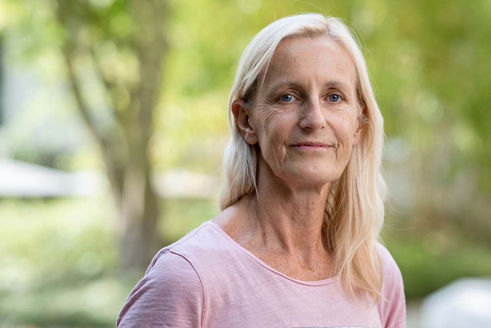
Using imaging techniques, in particular electron microscopy, Prof. Dr. Jacomina Krijnse Locker studies how viruses reprogram their host. In the process, she looks for targets to eliminate the pathogen.
How do viruses multiply? This question many people may have asked during the Corona pandemic. Those looking for in-depth answers quickly realize that science cannot explain it down to the last detail yet.
Viruses hijack the host cell, that much is certain. Equipped with little genetic material they manage to manipulate the cell such that it produces and releases masses of new viruses. Host cell membranes play a role in this process, as does the cytoskeleton, structures that provide cells with a frame. But what happens exactly in the host cells during infection? How do the multiplication strategies of viruses differ? Can specific steps of the viral replication cycle be stopped, thus offering a therapeutic approach?
Viruses: “Fascinating and terrifying”
Prof. Dr. Jacomina Krijnse Locker has been working on such research questions since the 1990s. She addresses these by directly observing viruses in their host cells: under the electron microscope (EM), with advanced light microscopes, and with the combination of both, correlative light- and electron microscopy (CLEM). The way viruses manipulate their host is both “Fascinating and terrifying”, she wrote in a recent editorial for the journal Molecular Microbiology. The SARS-CoV-2 pandemic proved that again, underscoring the need for fundamental research in the virus field.
The 64-year-old electron microscopy expert originates from the Netherlands. Her first professional position, in the faculty of veterinary medicine in Utrecht, led her to work on corona viruses in the research group of Prof. Peter Rottier. “In those days, hardly anyone outside of veterinary medicine was interested in corona viruses,” reports the head of the “Electron Microscopy of Pathogens Research Group” at the Paul Ehrlich Institute (PEI) in Langen near Frankfurt am Main. “They caused severe disease in animals, such as pigs, cats, mice, chicken and were considered harmless common-cold viruses in humans.” Her early expertise in Corona viruses is as useful to Krijnse Locker’s work today as her knowledge of vaccinia virus – the vaccine strain that lead to the eradication of smallpox, flaviviruses, several of which cause serious diseases in tropical countries, or the HI virus.
The challenge begins with sample preparation
In electron microscopy (EM), experience gives you a head start. The aim is to generate scientifically meaningful images of particles much smaller than cells – including viruses. In EM the scientific challenge begins long before the actual imaging of the cell’s interior. It lies in the preparation of the sample.
The structures that researchers want to see have to be properly identified in the first place – in a microworld where less than a nanometer (a millionth of a millimeter) makes huge differences. They should not be destroyed or altered by the preparation for EM. After all, scientists want to observe the processes in the host cell as close to reality as possible without preparation artifacts.
Viral pathogens of tropical diseases in comparison
Finding the right conditions for the preparation of samples is sometimes very time-consuming. Weeks and months can pass before something appears on the screen of an EM that can be scientifically evaluated. In the projects funded by the LOEWE center DRUID, Jacomina Krijnse Locker and her team are working on a special challenge: In cooperation with other DRUID groups, they study various viruses that cause tropical diseases that so far cannot be treated. Among these are flaviviruses that Krijnse Locker has worked on previously, such as Zika virus, West Nile virus, Dengue virus, as well as the Bunyavirus Rift Valley Fever virus.
Her team includes two technical assistants, two postdoctoral fellows and two graduate students. “We are trying to establish routine procedures as a basis for studying different viruses,” Krijnse Locker explains. “Although we are already quite far, such working protocols will need to be adapted to different viruses and their respective questions.”
Important findings on SARS-CoV-2 during the pandemic
In 2020, Jacomina Krijnse Locker accepted the DRUID professorship “Neglected Infectious Diseases with a Focus on Imaging” at the University of Giessen together with the PEI according to the so-called Jülich Model. She works with her group at PEI and teaches in parallel at the University of Giessen in the Department 08 of Biology and Chemistry. Only 6 weeks after her start, her lab was hit by the first Coronavirus lockdown. This is where her Corona expertise paid off; rather than staying at home Krijnse Locker successfully isolated and purified SARS-CoV-2 and together with collaborating teams found out important details about the spike protein.
Working in an EM lab requires a steady hand and a lot of endurance. After preparing the samples, the next challenge is to correctly interpret what the images on the monitor mean; “Is that really a membrane structure around a virus, or does it belong to the host cell?” Is it possible that you are looking at something that has nothing to do with the research question? If something important is seen in the microscope, the observations need to be checked and validated- using other microscopic methods, by quantifying the observations and analysing the images. From the time a single image is captured it is still a long way to better understand disease-causing viruses.
Dutch special path to research career
Jacomina Krijnse Locker says she immediately became excited about imaging the first time she sat in front of a microscope. Before she already realized that she wanted to work with pathogens. Right after graduating from high school, it wasn’t so clear.
“I didn’t really know what I wanted and first completed a study to become a technical assistant,” she reports. However, already during her first position in a research laboratory she realized that she wanted to do research. After several years of technical assistant, she went on to do her PhD on corona viruses at the University of Utrecht, without university degree which is possible in the Netherlands. After her PhD she worked at the “European Molecular Biology Laboratory” (EMBL) in Heidelberg and habilitated at the University of Heidelberg, where she subsequently headed the Electron Microscopy Core Facility. From 2015 to 2019, she was responsible for the Electron Microscopy Unit at the Institut Pasteur in Paris.
Germany, she says, “offers a very good environment for electron microscopy.” There is plenty of state-of-the-art equipment and the appropriate expertise to keep each other up to date, she says. The DRUID project enables an open exchange, something that was missing in Pasteur, she says: “We find each other and cooperate in an uncomplicated way. It helps achieve DRUID’s overall goal: identifying potential targets and drugs to treat neglected tropical infectious diseases.
Many groundbreaking findings in virology are brand new
Observing viruses directly in the host cell has two sides, says Krijnse Locker: “It teaches us about the virus and about the cell.” This increases the prospects of finding medically relevant targets that destroy the viruses without harming the host. Currently, Jacomina Krijnse Locker focuses on how viruses leave their host, to enter and infect other cells.
As always in science, new findings immediately raise new questions. “How do viruses manipulate their host so that the steps the virus needs to multiply are as efficient as possible? So that everything takes place in the host cell at the right time and at the right place? And at the same time, how do viruses manage to ensure that the host cell does not die immediately?” summarizes Krijnse Locker. There is still much to discover in the electron microscope.
Contact
Prof. Dr. Jacomina Krijnse Locker
Elektronenmikroskopie von Pathogenen
Paul-Ehrlich-Institut
Paul-Ehrlich-Straße 51-59
63225 Langen
Tel.: +49 (0)6103-77 2011
E-Mail: Jacomina.KrijnseLocker(at)pei(dot)de

